What Does "Ultrasound Probe Testing" Mean in a Clinical Context?
"Ultrasound probe testing" refers to the performance and safety assessment of medical diagnostic ultrasound transducers — the hand-held devices that convert electrical energy into the acoustic pressure waves used to form a diagnostic image. It is not a single measurement. A complete evaluation combines image-quality quality assurance (QA) against tissue-mimicking phantoms, electrical safety testing to the IEC 60601 family, and acoustic-output measurement with hydrophones. The framework is anchored by three standards that each govern a different question: IEC 60601-2-37 (the particular safety standard for ultrasound transducers themselves), IEC 62353 (recurrent electrical-leakage and safety testing of medical electrical equipment), and IEC 62359 (acoustic-output parameter declaration). The reason the test matters clinically is straightforward: an array with one or more dead elements, a delaminated acoustic lens, or a stray leakage current can produce a passable-looking 2D image yet degrade diagnostic confidence — and in transesophageal (TEE), intraoperative and endocavitary probes, even small acoustic or electrical deviations can directly compromise patient safety.
Why Doesn't a Good Scanner Image Mean the Probe Is Healthy?
The most persistent misconception in probe testing is that a normal-looking image proves the transducer is fine. It does not. A degraded element may produce an acceptable image in 2D B-mode yet break down under depth or Doppler conditions; a skewed beam profile can slip past a casual scan review while distorting diagnostic data; and image quality alone does not reveal whether the probe is emitting the correct acoustic pressure or whether thermal risk has changed after a repair. This is the gap between calibration and validation: calibration confirms the probe outputs energy within a defined range; validation confirms the device performs safely and accurately in a clinical context. A probe may be acoustically calibrated but still emit non-uniform beams, or "look fine" while clinically relevant degradation persists. Competent probe testing therefore treats image review as one input among several, not as the verdict.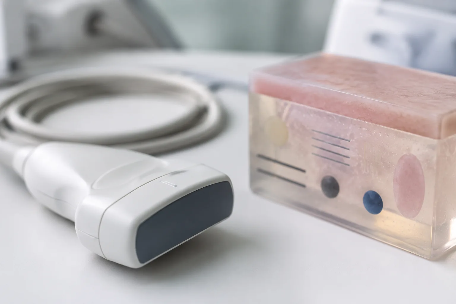
What Are the Six Field Tests That Define Image-Quality QA?
The industry-recognised field testing of image performance follows a defined set of functional tests established by the major accreditation boards — the American College of Radiology (ACR) and the American Institute of Ultrasound in Medicine (AIUM). These six tests, run on a tissue-mimicking phantom (e.g. Gammex or ATS) with image-processing options (harmonics, compounding, smoothing) switched off, give objective, repeatable pass/fail data:
- Image uniformity (element/channel testing) — element-to-element comparison across the full image width at 3–6 cm depth, single focal point in the near field. Ranking: 0 flaws = OK; 1–2 minor fine shadows = operational, monitor; ≥3 minor = borderline, replace soon; any major/wide shadow on multiple elements = remove from service immediately.
- Maximum depth of penetration — deepest vertical line target visualised; should stay constant over the probe's life. A 5% decrease from baseline is cause for concern; a 10% decrease triggers corrective action.
- Functional resolution — depth of the deepest, smallest anechoic target clearly visualised.
- Geometric accuracy — vertical distance within 1.5% or 1.5 mm (whichever greater); horizontal within 2% or 2 mm. Gross inaccuracy flags a system-level problem.
- Spatial resolution — minimum distance at which two targets are resolved axially (parallel to beam, depth-independent) and laterally (perpendicular, varies with depth/focus).
- Contrast resolution — ability to distinguish grey-scale targets of varying brightness; all shades must be clearly visualised with circular, graduated targets.
Two further tests — cable noise (flex strain reliefs in CW Doppler for cardiac or colour Doppler for non-cardiac probes, looking/listening for noise) and electro-mechanical functionality (3D/4D volumetric probes: apply slight dome pressure, stress strain reliefs; no error message) — are not in the accreditation core but complete a comprehensive assessment. The common thread is that these tests use identical presets every time — the same phantom model, the same scanner preset per probe type — so that a change between inspections reflects the probe, not the test conditions.
How Do You Catch Probe Defects Without Specialised Equipment?
Between formal phantom QA sessions, the most sensitive routine screen is the in-air reverberation test. The probe is switched on (using its usual clinical preset — e.g. abdominal preset for a curvilinear transducer) and held in air; the reverberation pattern appears as several bright, parallel-running lines in the near field. In a healthy probe these lines are parallel and homogeneous. The four classic defect signs are:
- Dropout — loss of continuity of the reverberation pattern, increasing distally; indicates piezo-element failure.
- Delamination — disrupted pattern without complete signal loss; lens or matching-layer separation, or a weak element.
- Non-uniformity — reverberation lines running non-parallel; implies image distortion, and warrants urgent replacement.
- Lens wear — subtle peripheral change in reverberation depth from long-term use; monitor for progression.
A refinement is the paper-clip method: a small reflective object (a paper clip) is moved along the lens surface with a drop of acoustic gel to accentuate smaller dropouts. A caveat — phased-array transducers are harder to assess this way because a single element failure impacts the whole scan field, which is why a modified paper-clip test using M-mode has been proposed for them. Visual inspection of the physical probe remains the daily first line: lens holes/cuts/bubbles/separation, housing cracks, strain-relief failure, cable cuts and roll-over damage, and bent/corroded connector pins each flag likely internal damage.
What Do the Three IEC Standards Actually Test For?
These standards are not interchangeable, and a complete report usually cites all three because they answer different questions:
- IEC 60601-2-37 — the particular standard for the ultrasound transducer itself. It sets the construction, marking and basic safety requirements a probe must meet as placed on the market. This is the standard relevant to type testing of a new or repaired probe.
- IEC 62353 — recurrent electrical safety testing (leakage current, protective-earth continuity, insulation) of medical electrical equipment in service. For intracavitary and TEE probes, where the device enters the patient, leakage testing is paramount and must be performed after any servicing.
- IEC 62359 — declaration of acoustic output parameters (mechanical index, thermal index, pressure fields). Hydrophone measurement of the acoustic pressure field is how a lab verifies that a probe still emits within the mechanical- and thermal-index thresholds — essential for high-risk probe categories and for FDA submission compliance.
Verifying all three together is what separates a probe that merely produces an image from one that is clinically and electrically safe.
Repair, Remanufacture and the FDA Distinction
A recurring theme in probe testing is the legal boundary between servicing and remanufacturing. Under FDA guidance, service activities that significantly alter a device's performance, safety, or intended use are remanufacturing and trigger additional regulatory controls — formal registration as a device manufacturer, a 510(k) or PMA, and full quality-system compliance under 21 CFR Part 820. The practical implication for testing: if a probe is repaired without quantitative documentation that it was returned to OEM specifications, its regulatory status becomes ambiguous and the liability shifts to the hospital and the healthcare technology management (HTM) team. This is why post-repair probe testing must produce traceable validation reports — electrical-leakage data, acoustic-output mapping, beam-uniformity analysis, and OEM-benchmarked image-performance metrics — not just a visual scanner check. The AIUM position is explicit: users should ensure that replacement and remanufactured transducers are cleared by the FDA per the relevant Guidance, and should obtain from the OEM a list of qualified third-party repair organisations.
FAQ
How often should ultrasound probes be tested?
Acceptance testing when a probe is new, returns from storage, or has been repaired/replaced; periodic QA at least annually per accreditation-board recommendation, or at OEM-recommended intervals (whichever is sooner); and daily visual inspection by sonographers. Probes aged 2+ years carry a markedly higher incidence of internal structural defects — published service data put it near 25% — so older units benefit from more frequent formal QA.
What is the in-air reverberation test and why do it?
It is a no-equipment screen: hold the energised probe in air and assess the near-field reverberation pattern. Healthy probes show parallel, homogeneous bright lines; dropout, delamination, non-uniformity or lens wear indicate element or lens faults. It detects a large proportion of equipment damage and should be documented at regular intervals for every probe in use.
What counts as a failing result on penetration depth?
Compared to the probe's baseline measurement, a 5% decrease in maximum depth of penetration is cause for concern; a 10% decrease requires corrective action to the probe or scanner. Root causes include acoustic-lens damage, fluid/gel infiltration, matching-layer damage, or array degradation over time (typical beyond 8–10 years of service).
Why is electrical leakage testing especially important for TEE and endocavitary probes?
These probes enter the patient's body — oesophagus, vagina, rectum, or surgical field — so any leakage current is delivered directly to tissue or mucosa. IEC 62353 leakage testing after any servicing (and a pneumatic leakage test on the TEE insertion tube) is therefore non-negotiable for these categories, where even small electrical or acoustic deviations carry the highest clinical risk.
Does a normal scanner image mean a repaired probe is validated?
No. A normal image confirms visual functionality, not that the probe meets OEM acoustic, electrical or image-performance tolerances. Validation requires quantitative data — hydrophone acoustic-output mapping, leakage testing, phantom-based image metrics — benchmarked against OEM specifications. Without it, the repair may legally constitute undocumented remanufacturing.
Our Testing Capabilities
As an ISO/IEC 17025-accredited third-party laboratory, Beijing ZKGX Research provides ultrasound probe testing aligned to the IEC 60601 family and accreditation-board QA frameworks:
- Image-quality QA on tissue-mimicking phantoms: image uniformity (element/channel ranking), maximum depth of penetration and functional resolution (with 5%/10% drift flags), geometric accuracy (vertical ≤ 1.5%/1.5 mm, horizontal ≤ 2%/2 mm), spatial resolution (axial/lateral) and contrast resolution.
- Electrical safety to IEC 62353 (leakage current, protective-earth continuity, insulation), with particular attention to intracavitary, TEE and intraoperative probes.
- Acoustic-output verification with NIST-traceable hydrophone measurement (mechanical/thermal index, pressure-field mapping) per IEC 62359, and probe type testing to IEC 60601-2-37.
- Physical and defect screening: in-air reverberation and paper-clip methods, lens/housing/cable/connector visual inspection, cable-noise and electro-mechanical (3D/4D) checks, and root-cause mapping of visual defects to internal faults.
- Post-repair / remanufacture validation reports: traceable, OEM-benchmarked electrical, acoustic and imaging data to support FDA-equivalent compliance and audit readiness.
For related electrical-equipment conformity work, see our IEC laser equipment testing. If you have a specific probe model, clinical category (TEE / endocavitary / intraoperative / point-of-care) or compliance target, contact the laboratory to confirm the exact test set, standards and reporting format.